‘Mini-lungs’ reveal early stages of Covid-19 infection
Infecting mini lab-grown lungs with coronavirus may reveal how the virus damages the respiratory system and make it easier to fight the disease.
The main target tissues of SARS-CoV-2, the virus that causes Covid-19, especially in patients that develop pneumonia, appear to be alveoli.
These are the tiny air sacs in our lungs that take up inhaled oxygen and exchange it with carbon dioxide to exhale.
To better understand how the virus infects the lungs and causes disease, a team of scientists from the UK and South Korea turned to organoids – mini-organs grown to mimic real life organ behaviour.
They used tissue donated to Addenbrooke’s Hospital and Royal Papworth Hospital in Cambridge and Seoul National University Hospital to extract a type of lung cell known as alveolar type 2 cells.
Based on our model we can tackle many unanswered key questions, such as understanding genetic susceptibility to SARS-CoV-2
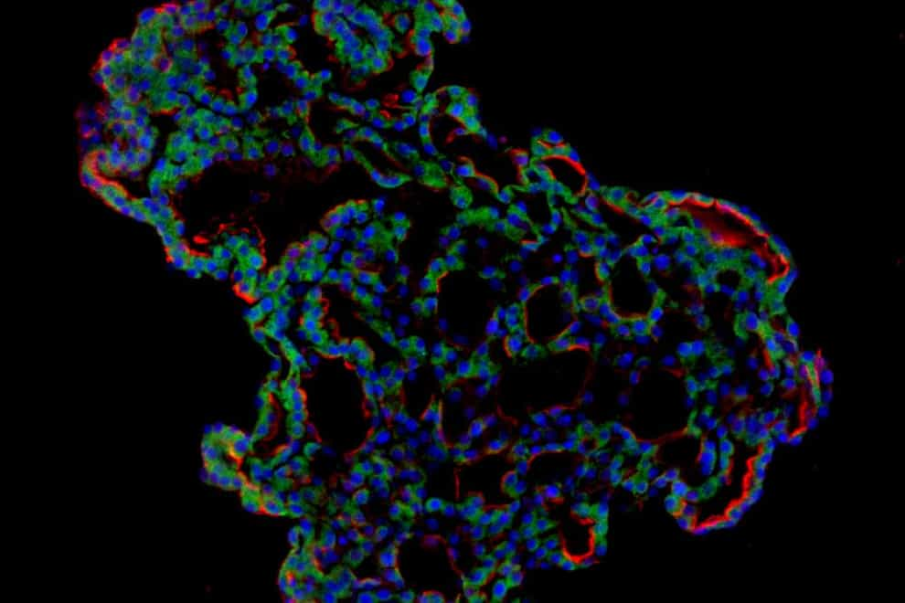
The researchers reprogrammed these cells so that they could grow into alveolar-like 3D structures that mimic the behaviour of key lung tissue.
Dr Joo-Hyeon Lee, co-senior author, and a group leader at the Wellcome-MRC Cambridge Stem Cell Institute, University of Cambridge, said: “We still know surprisingly little about how SARS-CoV-2 infects the lungs and causes disease.
“Our approach has allowed us to grow 3D models of key lung tissue – in a sense, ‘mini-lungs’ – in the lab and study what happens when they become infected.”
The organoids were infected with a strain of SARS-CoV-2 taken from a patient in South Korea who was diagnosed with Covid-19 on January 26 after traveling to Wuhan, China.
Researchers were able to study how the cells responded to the virus using a combination of fluorescence imaging and single cell genetic analysis.
They found that when the 3D models were exposed to SARS-CoV-2 the virus began to replicate rapidly and reached full cellular infection six hours after infection.
Around the same time, the cells began to produce interferons – proteins that act as warning signals to neighbouring cells, telling them to activate their antiviral defences.
According to the study, published in the journal Cell Stem Cell, after 48 hours the interferons triggered an innate immune response – the organoid’s first line of defence – and the cells started fighting against infection.
Sixty hours after infection a subset of alveolar cells began to disintegrate which led to cell death and damage to the lung tissue, the researchers found.
Clinical symptoms do not usually occur as quickly as the lung cell changes were observed in this experiment. Symptoms can sometimes take more than ten days after exposure to appear.
The scientists say there are several possible reasons for this. For example, it may take several days for the virus to reach the alveoli after infiltrating the upper respiratory tract.
They suggest it may also require a substantial proportion of alveolar cells to be infected or for further interactions with immune cells – resulting in more inflammation – before a patient shows symptoms.
Dr Young Seok Ju, co-senior author, and an associate professor at Korea Advanced Institute of Science and Technology, said: “Based on our model we can tackle many unanswered key questions, such as understanding genetic susceptibility to SARS-CoV-2, assessing relative infectivity of viral mutants, and revealing the damage processes of the virus in human alveolar cells.
“Most importantly, it provides the opportunity to develop and screen potential therapeutic agents against SARS-CoV-2 infection.”
Dr Lee added: “We hope to use our technique to grow these 3D models from cells of patients who are particularly vulnerable to infection, such as the elderly or people with diseased lungs, and find out what happens to their tissue.”
The best videos delivered daily
Watch the stories that matter, right from your inbox